Steven Salzberg Publishes Reanalysis of UCSF's Defective Lancet Paper in F1000
After Yoav Gilad’s reanalysis of mouse ENCODE data, respected computational biologist Steven Salzberg took the F1000 route to critically re-examine the claims of another high-profile paper.

Over the last few years, bio-medical journals have seen severe outbreak of high-profile papers of poor quality. Cleaning up the mess through the same exclusive journals is extremely difficult (check the experiences of Dan Graur or Lior Pachter), but thanks to alternate publishing venues, there is finally some hope.
Today’s paper under fire is about the recent enterovirus D68 outbreak that caused ‘mystery paralysis’ and polio-like symptoms. The authors claimed to have found (A novel outbreak enterovirus D68 strain associated with acute flaccid myelitis cases in the United States from 2012-2014: a retrospective cohort study) through metagenomic sequencing -

48 patients were included: 25 with acute flaccid myelitis, two with enterovirus-associated encephalitis, five with enterovirus-D68-associated upper respiratory illness, and 16 with aseptic meningitis or encephalitis who tested positive for enterovirus. Enterovirus D68 was detected in respiratory secretions from seven (64%) of 11 patients comprising two temporally and geographically linked acute flaccid myelitis clusters at the height of the 2014 outbreak, and from 12 (48%) of 25 patients with acute flaccid myelitis overall. Phylogenetic analysis revealed that all enterovirus D68 sequences associated with acute flaccid myelitis grouped into a clade B1 strain that emerged in 2010. Of six coding polymorphisms in the clade B1 enterovirus D68 polyprotein, five were present in neuropathogenic poliovirus or enterovirus D70, or both. One child with acute flaccid myelitis and a sibling with only upper respiratory illness were both infected by identical enterovirus D68 strains. Enterovirus D68 viraemia was identified in a child experiencing acute neurological progression of his paralytic illness. Deep metagenomic sequencing of cerebrospinal fluid from 14 patients with acute flaccid myelitis did not reveal evidence of an alternative infectious cause to enterovirus D68.
Salzberg found several defects in their analysis. The authors did not use any standard bioinformatics program and instead reinvented their version of square wheel, which they published as a separate bioinformatics paper. Unfortunately, those great programs failed to find out that two patients had other major bacterial infections (Haemophilus influenzae and severe Staphylococcus aureus). Moreover, the authors did not remove human DNA from patients’ data before public submission, as they claimed to have done likely to protect patients’ privacy. The full abstract of Salzberg is shown below -
Metagenomic sequence data can be used to detect the presence of infectious viruses and bacteria, but normal microbial flora make this process challenging. We re-analyzed metagenomic RNA sequence data collected during a recent outbreak of acute flaccid myelitis (AFM), caused in some cases by infection with enterovirus D68. We found that among the patients whose symptoms were previously attributed to enterovirus D68, one patient had clear evidence of infection with Haemophilus influenzae, and a second patient had a severe Staphylococcus aureus infection caused by a methicillin-resistant strain. Neither of these bacteria were identified in the original study. These observations may have relevance in cases that present with flaccid paralysis because bacterial infections, co-infections or post-infection immune responses may trigger pathogenic processes that may present as poliomyelitis-like syndromes and may mimic AFM. A separate finding was that large numbers of human sequences were present in each of the publicly released samples, although the original study reported that human sequences had been removed before deposition.
Thankfully, the main claim of the original paper is still valid as Dr. Salzberg pointed out in a private email.
I should add that the main finding of Greninger et al. is not really in doubt - their primary analysis showed that the 2014 strain of enterovirus D68 formed a new clade. But the secondary analyses included some surprising flaws.
-——————————-
Edit.
1. Omics!Omics! blog wrote -
Leaky clinical metagenomics pipelines are a very serious issue
-——————————-
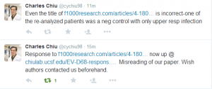
2. Charles Chiu, the senior author of UCSF paper, posted a response on his website signed by three authors (incl. him). I am quoting it here, but please feel free to discuss at the F1000 site.
http://chiulab.ucsf.edu/EV-D68-response.docx
This manuscript raises two main criticisms of our paper in their re- analysis. Here we directly address the 2 main points:
1. The authors claim that bacterial reads were seen in the nasopharyngeal / oropharygneal swab (NP/OP) metagenomic data that were “missed” in the original study. They were able to assemble two genomes, from Haemophilus influenzae and methicillin-resistant Staphylococcus aureus. We would like to highlight that these reads and bacteria were not missed but instead we did not discuss the bacterial/fungal portion of the NP/OP swabs in the manuscript due to difficulties in clinical interpretation.
As quoted from the supplementary section of our manuscript published in Lancet Infectious Diseases, with our emphasis in underline:
Lancet ID Supplementary Material: NGS libraries constructed from NP/OP samples were treated with DNase following nucleic acid extraction to reduce background from the human host and bacterial flora. As this protocol reduces sensitivity of detection and speciation for non-viral microbes (i.e. bacteria, fungi, and parasites), only viral sequences are shown for the NP/OP samples. The ability to detect DNA viruses is also impacted by the use of DNAse and we cannot exclude the possibility that our data is biased by reduced sensitivity for detection of DNA viruses.
a) The authors also state that we “did not report finding any bacterial reads from this sample” but that does not mean that we did not detect any bacterial reads. In fact, we detected both the Haemophilus influenzae and methicillin- resistant Staphylococcus aureus reported by the authors:
The number of bacterial reads found in all metagenomic samples analyzed is shown in Supplementary Table 3, Column N. Bacterial reads for the two samples in question are listed as US/CA/09-871: 5,614,487 bacterial reads and US/CA/12-5837: 28,676,383 bacterial reads.
In the main text we also state that the purpose of metagenomic NGS on NP/OP samples was to “to aid in the recovery of enterovirus D68 genome sequences and detect potential co-infections from other viruses”.
b) In addition, although we reported the presence of bacterial reads in the NP/OP data, detected using SURPI/SNAP, we did not discuss this in the paper due to a number of reasons:
Our focus on looking for CSF (bacterial viral fungal and parasite) pathogens in the setting of AFM our clinical perspective that the presence of bacterial reads in NP/OP swabs from children most often reflect colonization / carriage and not infection (see http://www.biomedcentral.com/1471-2180/10/59, http://www.ncbi.nlm.nih.gov/pubmed/15999003, http://www.ncbi.nlm.nih.gov/pmc/articles/PMC3962756/, http://www.ncbi.nlm.nih.gov/pubmed/12394812, etc. for papers on nasal carriage of Staphylococcus and Haemophilus in healthy children)
our treatment of the nucleic acid extracts with DNase to help reduce host background, which would bias accurate metagenomic interpretation and the accurate counting of bacterial and fungal reads since this procedure degrades bacterial / fungal genomic DNA due to high levels of respiratory tract colonization, NP/OP samples nearly always contain bacterial sequences that will constitute the majority of the non-human reads and beyond reporting read numbers, we did not comment on this in the paper.
c) Of note, the sample in question with Haemophilus influenzae (US/CA/09-871) came from a 2009 case of upper respiratory infection alone. This subject did not have either encephalitis or acute flaccid myelitis (Table 2 and results in Greninger et al.). The statements in the manuscript that “This patient was reported as having encephalitis and severe respiratory illness…” and “the sequence evidence here suggests that the patient might have had complications from H. influenzae-associated encephalitis or encephalomyelitis…” as well as the title of the manuscript are therefore incorrect.
2. The authors point out that there are residual human reads in the deposited data.
We acknowledge that we have been using SNAP, which is a global aligner, to extract out human reads for our SRA submissions. This algorithm will miss human reads because of low-complexity sequences at the ends, residual adapters, etc. We agree that we probably should have used a local aligner such as BLASTn at a low threshold level to more completely extract out human reads, or a k-mer approach such as Kraken. We appreciate the authors point on the importance of clearing human sequences from metagenomic data and will plan on resubmitting this more completely filtered data to the SRA in the near future.
The re-analysis presented here gives the erroneous impression that bacterial colonization has a strong association with the devastating acute flaccid myelitis (AFM) syndrome seen in our patients. As we note above, Haemophilus influenzae was found in a control patient with no neurological symptoms, and is thus not relevant to AFM. While MRSA colonization may possibly be a factor in AFM, it is extremely unlikely given its detection is a single case and the fact that MRSA nasal carriage in healthy children is well-described. We should also point out that bacterial cultures of cerebrospinal fluid (CSF) from all of the patients in the study were negative and that metagenomic next- generation sequencing did not reveal any evidence of bacterial infection in the central nervous system (CNS).